Rosalind Franklin was an English biophysicist and geneticist who made tremendous contributions to the field of DNA research and its applications in medicine. Unfortunately, her recognition lagged behind that of her male colleagues, who took credit for her work. Today, she is largely remembered because of their mistreatment of her. Franklin’s early education followed a conventional path for girls in the 1930s, she attended grammar school, graduated from Newnham College at the University of Cambridge with a degree in natural sciences, and eventually received a PhD from again the University of Cambridge for her thesis on carbon chains as markers for biological activity in photosynthesis.
Basics of DNA Research
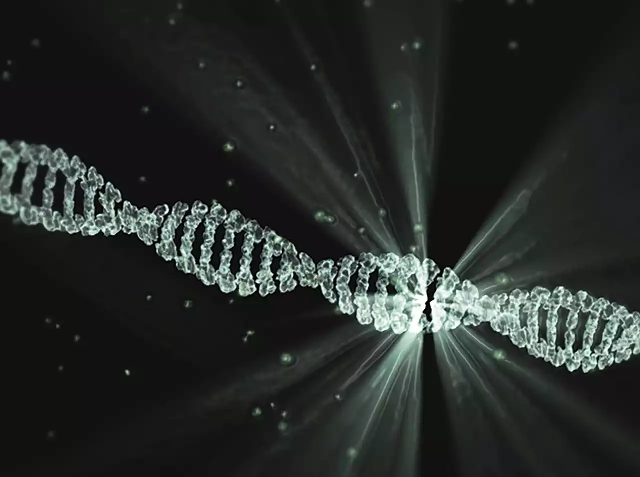
DNA (deoxyribonucleic acid) is a chemical found in almost every cell in the human body. Scientists believe that DNA carries genetic information, or instructions, that help control the way our bodies work. The DNA in our cells is organized into structures called chromosomes. Many chromosomes exist inside each cell, and each chromosome is made up of many genes. Genes are regions of DNA that contain instructions about how the body grows and develops. Most genes are found on only one chromosome, but a few genes are found on more than one chromosome. Genes are considered the basic units of heredity. Scientists use the word “DNA” as shorthand for “genes” or “chromosomes.”
X-ray diffraction and DNA research
When the relevant molecular biological techniques were developed by the mid-1940s, DNA proved largely intractable to these. That changed in 1948, when Rosalind Franklin and Maurice Wilkins, who worked at King’s College London, were able to produce X-ray diffraction images of DNA fibers with a density close to that of water. Such images were possible only because Franklin had recently developed a method of producing extremely thin fibers of DNA, which she had demonstrated to Wilkins. This breakthrough was the culmination of lengthy efforts by both researchers to investigate the structural aspects of the molecule. Wilkins had been interested in DNA since the late 1930s when he had begun collaborating with Franklin. Her research on the structure of coal had brought her into contact with techniques of X-ray analysis, which she evidently found applicable to her studies of the molecular structure of DNA. For many years, Franklin and Wilkins had tried to produce good X-ray images of DNA fibers, and failed. But, as Franklin herself noted, “A fresh look at a difficult X-ray problem often leads to a sudden solution.” This occurred in early 1948 when she and Wilkins were able to produce images of very high quality.
Continued Research on DNA
Franklin’s ability to produce high-quality images of DNA enabled her to explore a variety of aspects of the molecule’s structure. She was able to confirm that the molecule was shaped like a helix and to explore the number and twist of the DNA helices in the fibers. She was also able to explore the spatial arrangement of the bases within the helices. At this point, however, Franklin’s research on DNA came to an abrupt halt. This was because she had decided to change fields and study the tobacco mosaic virus (TMV), a pathogen that caused disease in plants. This decision was unusual because at the time most biologists focused on the study of protein molecules and TMV contains no proteins.
Double helix discovery and Rosalind Franklin’s role
In 1951, two biologists, James Watson and Francis Crick began working at the Cavendish Laboratory in Cambridge, where Franklin had once worked. Crick had learned that Franklin was no longer doing research on DNA and had decided to investigate the problem. It soon became clear to Crick and Watson that the best way to establish the structure of DNA was to construct a double helix. Franklin’s X-ray images of DNA fibers had shown that the molecule had a helical structure, but she had not been able to determine the positions of the bases within the helix. Crick and Watson, on the other hand, had ample experience with X-ray diffraction and were able to construct a double helix of DNA.
A new perspective on Rosalind Franklin
The discovery of the double helix of DNA created a sensation among biologists. Researchers realized that the structure explained many of DNA’s biological properties, including its capacity to instruct cells to grow and reproduce. Although Crick and Watson received most of the recognition for this discovery, Franklin’s contribution was not overlooked. Indeed, Watson, in his first book about the discovery of the structure of DNA, emphasized Franklin’s role, he wrote that her X-ray images were “the most beautiful ever taken.” Yet Watson’s wording also suggested that Franklin had not been given ample credit for her efforts. This view was reinforced by Franklin’s family and associates, who expressed outrage at the slighting of her contributions. By the late 1970s and early 1980s, as Watson’s account of the discovery of the structure of DNA was increasingly accepted, many who had known Franklin came to think that she had been badly treated.
Final Words
Franklin’s contributions to the discovery of the structure of DNA should not be minimized. She was the first to provide X-ray images that clearly indicated the helical structure of the molecule. She also made important contributions to the methods used in producing X-ray images and, after 1953, when she turned to the study of viruses, she made significant discoveries in this field as well. As a result of her efforts, it is now clear that Franklin was one of the most important scientists of the 20th century. But, because she died in 1958, many people are unaware of her achievements. It is true that Franklin lacked the self-promoting skills of her male colleagues. But her fate can also be understood in terms of the expectations that others brought to their encounters with her. Franklin’s excellence in the laboratory was clear to all who saw her work and was evident in her images. From the start, she was recognized as a scientist of unusual ability.

 Automobiles – The invention of the Pneumatic Tire
 Samsung Phones and Android System
 Power Banks For Mobile Phones
 Louis Pasteur, a Pioneer in Microbiology
 Francis Crick, an Inspirational Scientist
 Charles Darwin and His Famous Work
 Alexander Fleming and the Discovery of Penicillin